Projects
The research program in the Allbritton Lab is a multidisciplinary effort that brings to bear principles and techniques from chemistry, physics, engineering, and materials science to develop new assays and technologies for biomedical applications. The ongoing work in the lab comprises three major focus areas:
- Analytical techniques for single-cell biochemical assays
- Microfabricated platforms for sorting and cloning cells
- Microengineered systems for recapitulating organ level function.
Many of the lab’s projects and extensive collaborations within these focus areas involve the development and application of new technologies for oncology and stem cell research.
Our research projects
Chemical Cytometry
Analytical Techniques for Single-Cell Biochemical Assays
The advent of effective pharmacologic compounds targeting specific signaling pathways for clinical applications has created a demand for enzymatic assays that can be performed on single cells in disease models and patient samples. Biochemical analyses of aberrant signaling pathways are now needed to direct the best treatment option as well as to assess treatment efficacy in individual patients. The goal of this research effort is to produce the instrumentation and chemical tools needed to directly assess the catalytic activity of kinases and other signaling molecules in living cells from disease models and cancer patients.

The lab has a long history in developing novel technologies using highly sensitive analytical methods including capillary- and microfluidic-based electrophoresis. Current research efforts are directed at the design and development of biochemical substrates using peptide and lipid chemistry to create reporters of cellular signal transduction. Additionally, single-cell preservation techniques to improve time resolution and analyte retention in downstream analysis are being investigated. Instrumental advances in the areas of high-throughput single-cell analysis and lab-on-a-chip platforms for cell-based assays are also an active area of research and development. The ability to identify multiple aberrant signaling pathways simultaneously in a patient’s tumor would enable individualization of multiple targeted therapies for that patient and offer the hope of developing individualized and improved therapies to supplant the existing standard of care.
Microraft Arrays
Microfabricated Platforms for Sorting and Cloning Single Cells
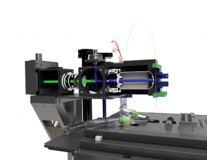
The lab has pioneered the development of novel microfabricated cell-array platforms to enable the analysis and isolation of cells while they remain on a culture substrate. Cells can be monitored at single or multiple time points and selected based on a wide range of criteria including morphology, fluorescence signatures, subcellular attributes, growth properties, and other time-dependent characteristics. These arrays are produced via dewetting methods to form molded rafts on a poly(dimethylsiloxane) base. Individual rafts are s electively detached employing a microneedle.

These arrays can be made in almost any size; for instance, millimeter-sized arrays for use with total sample sizes of hundreds to a few thousand cells (e.g. needle biopsies), to arrays of many centimeters with millions of culture sites for high-throughput screening of rare cell populations (e.g. circulating tumor cells). Numerous biomedically relevant applications of these microfabricated cell arrays are currently being pursued, including efficient cloning of murine embryonic stem cells for improved methods to create genetically engineered animals, isolation of cytolytic T lymphocytes based on their ability to kill tumor cells, and purification of circulating tumor cells and cancer stem cells from patient samples.
Organ-on-a-Chip
Microengineered Systems for Recapitulating Organ Level Function
The ability to monitor and control the environment at the cellular and tissue level is one of the most promising applications for microengineered systems. “Organ-on-a-Chip” platforms provide exquisite control of experimental variables, yet recapitulate much of the physiology of an intact organ. The lab is pursuing development and application of microTAS devices to recapitulate the microenvironments of the intestine.


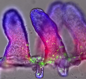
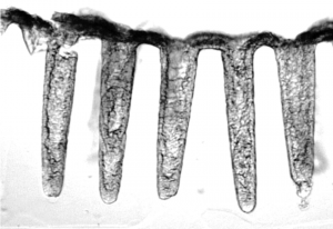
Purified intestinal stem cells or intestinal crypts are used to re-create colons on shaped arrays and contain the differentiated cell lineages as well as stem cells. Methods to create gradients across these tissues are being developed to mimic the chemical gradients thought to exist in vivo. The intestinal fluidic chips will make it feasible to combine cellular components from different genetically engineered mice for fundamental studies of intestinal biology, use in drug screening, and applications in toxicology.

